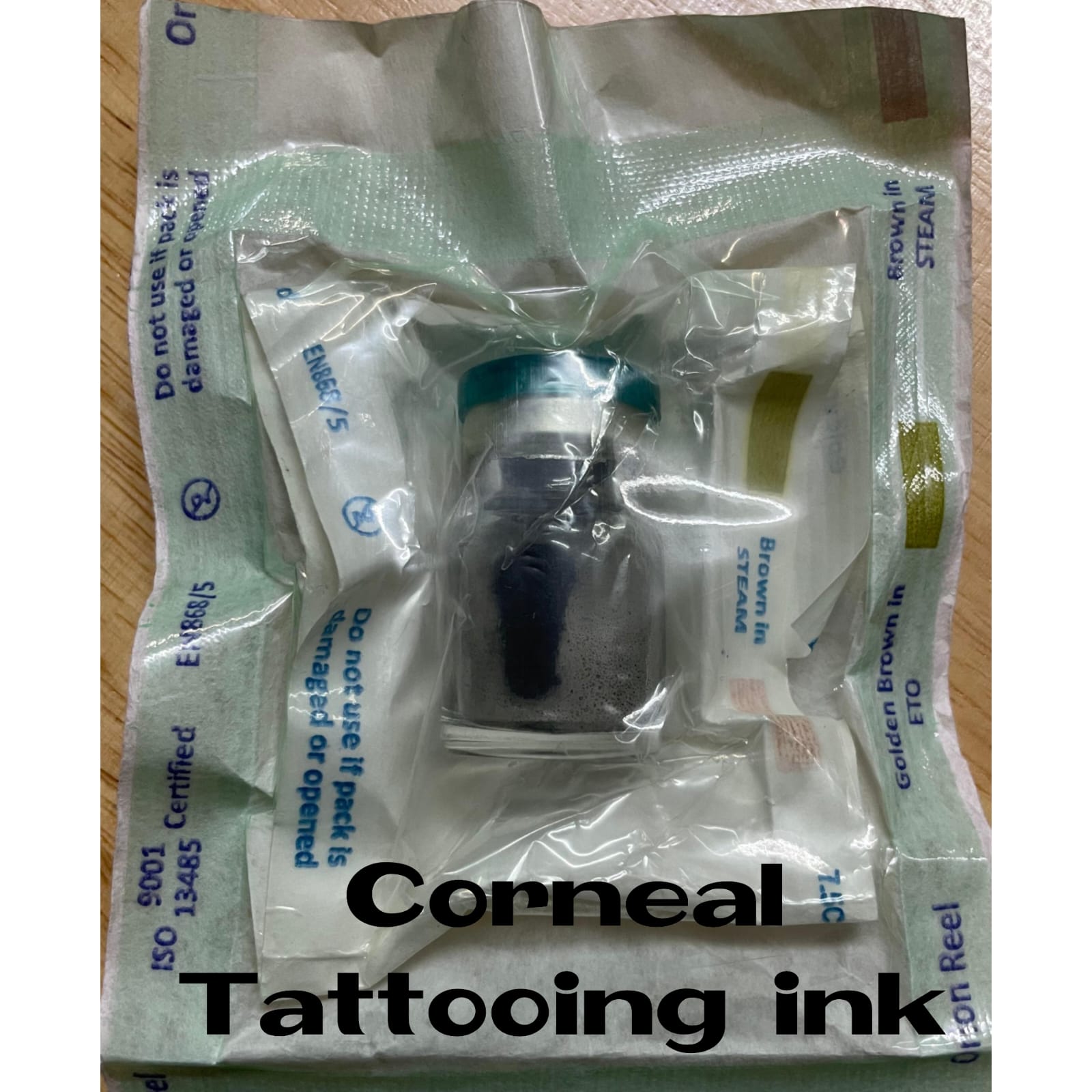
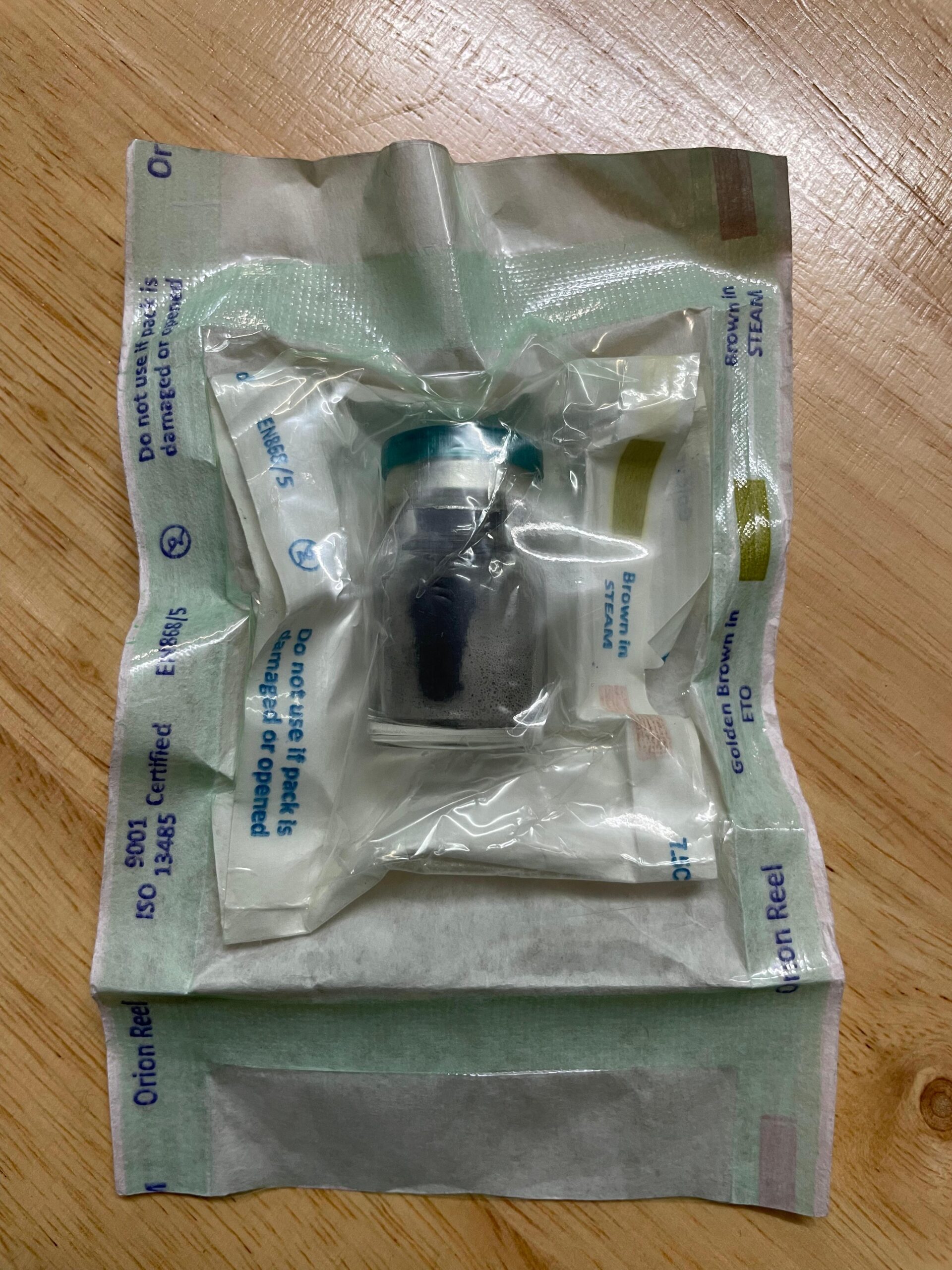
Description
Tattooing was known & practiced long before the Christian era. In primitive civilizations tattooing distinguished men of rank & status. Tattooing of the cornea for unsightly leucomas is of ancient lineage. In the practice of Ophthalmology you can’t always give vision to a patient. There are occasions when you need to add just a cosmetic angle in patient’s life, like in blind eye with corneal opacity one answer is tattooing. Then why tattooing is not done frequently by the ophthalmologists? There are two main reasons. Firstly there is non-availability of chemical pigment & secondly water pigments are used for tattooing are irritable to eye. Old water based pigment do not remain stable & in time they fade off rapidly. The ideal pigment for ocular tattooing should be inert and easily available & easy answer. It is not this new pigment for corneal tattooing is an easy answer. It is water-soluble but lipid soluble. It was tried initially on dyes & later water-solubles were prepared to get enneucleated afterwards. Next, the pigment was tried in patients of corneal opacity. The results are quite satisfactory.
TATTOOING VIDEO LINK
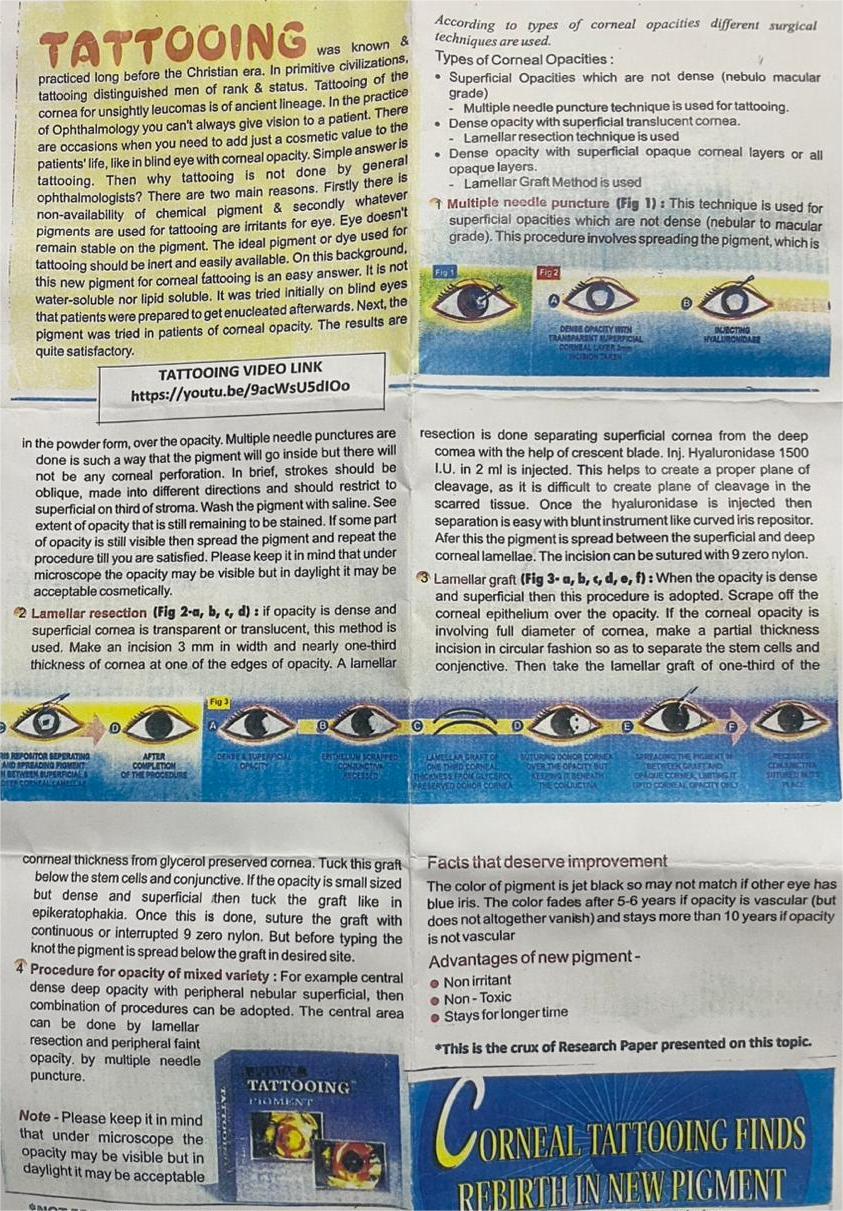
According to types of corneal opacities different surgical techniques are used.
Types of Corneal Opacities:
● Superficial Opacities which are not dense (nebulo macular grade)
➝ Multiple needle puncture technique is used for tattooing.
➝ Deep opacity with superficial translucent cornea.
● Lamellar resection technique is used.
● Dense opacity with superficial opaque corneal layers or all opaque layers.
➝ Lamellar Graft Method is used
1. Multiple needle puncture
This technique is used for superficial opacities which are not dense (nebular to macular grade). This procedure involves spreading the pigment, which is in the powder form, over the opacity. Multiple needle punctures are made in such a way that the pigment will go inside the but there is no corneal perforation. In brief, strokes should be oblique, made into different directions and should not be to the superficial third of stroma. Washing the pigment is used to spread onto graft so it will remain behind to be stained. Depth of opacity is still less so this is the simplest method which must be done till you are satisfied. Please be patient. It may hurt but cooperative main stays of good results. Done in by daylight timing in daylight.
⸻
2. Lamellar resection
If opacity is dense and superficial cornea is transparent or translucent, then this method is used. Make an incision 3 mm in width and nearly one-third thickness of cornea at one of the edges of opacity. A lamellar resection is done separating superficial cornea from the deep cornea with the help of crescent blade. Inj. Hyaluronidase 1500 I.U. in 2 ml is injected. This helps to create a proper plane of cleavage, as it is difficult to find the plane of cleavage in a scarred tissue. Once the hyaluronidase is injected, then separating the layer with instrument like curved iris repositor. Now spread the pigment behind lamellar resected surface of cornea. Suture the incised wound with 9 zero nylon.
⸻
3. Lamellar graft
When opacity is dense involving full diameter of cornea, make a partial thickness incision in circular fashion so as to separate the stem cell & conjunctive. Then take the lamellar graft of one-third of the corneal thickness from glycerol preserved cornea. Tuck this graft below the stem cells and conjunctive. If the opacity is small sized but dense and superficial then cut the graft like in conjunctiva. Once this is done, suture the graft with continuous or interrupted 9 zero nylon. But before giving final knot the pigment is spread below the graft in desired site.
⸻
4. Procedure for opacity of mixed variety:
For example, central dense deep opacity with peripheral nebular superficial, then combination of procedures can be adopted. The central area can be done by lamellar resection and peripheral faint opacity by multiple needle puncture.
⸻
Note:
Please keep it in mind that under microscope the opacity may be visible but in daylight it may be acceptable.
⸻
Facts that deserve improvement
The color of pigment is jet black so may not match if other eye has blue iris. The color fades after 5-6 years if opacity is vascular (but does not altogether vanish) and stays more than 10 years if opacity is not vascular.
Advantages of new pigment
● Non irritant
● Non-Toxic
● Stays for longer time
⸻
This is the crux of Research Paper presented on this topic.